In a groundbreaking achievement, scientists have successfully engineered the inaugural functional 3D-printed brain tissue capable of growth and connection formation akin to authentic human brain tissue.
This impressive achievement, spearheaded by a team at the University of Wisconsin–Madison, offers neuroscientists a novel tool to explore communication among brain cells and various regions of the human brain. This breakthrough holds promise for advancing treatments for conditions such as Alzheimer’s and Parkinson’s disease.
Because we can print the tissue by design, we can have a defined system to look at how our human brain network operates,” says Zhang. “We can look very specifically at how the nerve cells talk to each other under certain conditions.
According to the researchers, to comprehensively study the health and diseases of the human brain networks, a dependable model of living human neural tissues is essential. They emphasize that animal models fall short in replicating the intricate complexity of the brain.
Printing functional human brain tissue poses significant challenges, with many 3D-printed tissues currently lacking adequate cell connections. The maturation of neurons while maintaining tissue structure integrity is crucial, and supporting cells such as astrocytes play a vital role in ensuring proper tissue function.
Past endeavors employed non-biodegradable scaffolds, hindering neural cell migration. In a departure from conventional vertical layering, the team opted for horizontal layering of neurons derived from induced pluripotent stem cells. These cells were situated within a softer ‘bio-ink’ gel, contrasting with previous techniques.
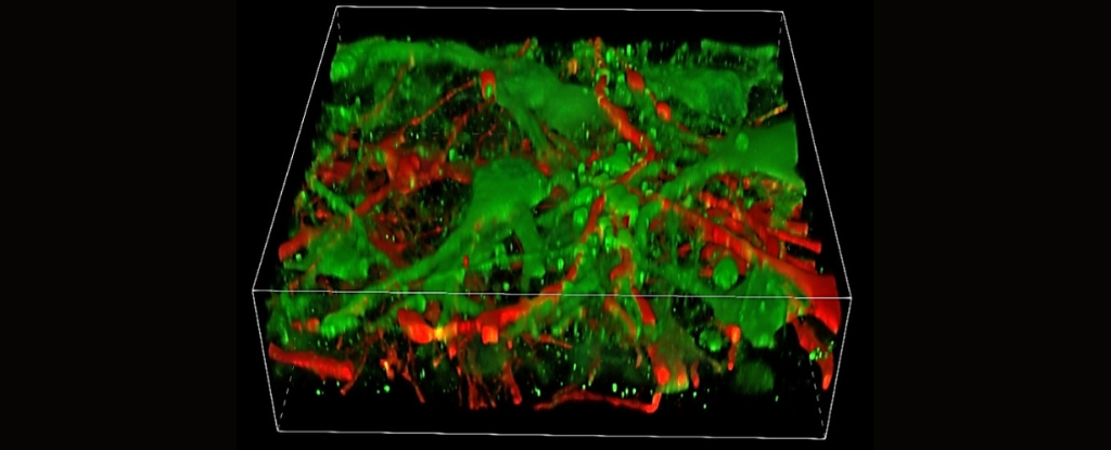
Within a few weeks, the cells in their printed tissue develop brain-like networks both within and across layers. Neurons engage in communication, signal transmission, neurotransmitter utilization, and establish networks, aided by additional supporting cells.
The tissue still has enough structure to hold together but it is soft enough to allow the neurons to grow into each other and start talking to each other,
explains Zhang.
Even when we printed different cells belonging to different parts of the brain, they were still able to talk to each other in a very special and specific way.
According to him, studying isolated components overlooks crucial aspects since the brain operates within networks. By printing brain tissue in this manner, it facilitates a more comprehensive observation of cell interactions.
We printed the cerebral cortex and the striatum and what we found was quite striking,
Zhang says.
Their research revealed that the axons extending in the printed brain tissue resemble the pattern found in the human brain, wherein cortical neurons project axons to the striatum.
This 3D-printing method offers precise control over cell types and arrangements, distinguishing it from brain organoids, which are miniature lab-grown organs utilized in brain research.
While the prototype lacks the ability to control the orientation of mature neurons and lacks the natural structure found in brain organoids, Zhang and colleagues assert that it serves as a valuable complement to organoids, offering an alternative approach to studying the brain under varied conditions.
It can be used to look at the molecular mechanisms underlying brain development, human development, developmental disabilities, neurodegenerative disorders, and more,
Zhang explains.
The team aims to enhance their process to produce more precise brain tissues with controllable cells.
The research findings have been released in Cell Stem Cell.